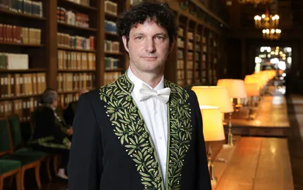
Présentation

Notre équipe étudie différents processus cellulaires : migration, régulation du volume et de la masse, division cellulaire. Nous sommes intéressés par le fonctionnement du cytosquelette, des organelles et de leur lien avec la méchanosensitivité. Nous développons des outils innovants basés sur des techniques de nano et micro-fabrication, afin de contrôler et moduler les principaux paramètres physiques et chimiques du micro-environnement cellulaire.
Ces outils sont couplés avec de la microscopie quantitative de haute qualité, et utilisés aux côtés de techniques de biologie moléculaire et cellulaire, afin d’obtenir une description quantitative des comportements cellulaires. Notre approche multidisciplinaire, en plus de mettre en lumière de nouveaux concepts basiques du comportement cellulaire, mène au développement de nouveaux outils avec un potentiel d’application dans la recherche biomédicale.
Nos recherches visent à comprendre comment les cellules prolifèrent et migrent lorsque l’espace est limité. Nous voulons comprendre comment les cellules (immunitaires et cancéreuses) peuvent produire un mouvement efficace dans des environnements confinés, et comment les contraintes physiques affectent l'enveloppe nucléaire et la division cellulaire.
Nos études sur la prolifération cellulaire sous contraintes externes ont été récompensées par un ERC Consolidator grant (2013-2018). Un ERC Synergy Grant (2023-2028), en collaboration avec les équipes d'Ana-Maria Lennon-Duménil, Giorgio Scita et Raphaël Voituriez, a également été obtenue afin d'étuider l'impact des formes cellulaires sur le comportement et le destin des cellules.
Matthieu Piel est l’auteur de plus de 352 publications, avec plus de 20000 citations (h-index 73). Il détient quatre brevets, et est co-fondateur de la société CYTOO, et de l’Institut Pierre-Gilles de Gennes pour la microfluidique.
M. Piel a également enseigné au Centre de Recherche Interdisciplinaire, et est actuellement enseignant en Biologie Cellulaire et en Biophysique dans plusieurs parcours de master à Paris.
M. Piel fut récompensé par la Médaille de Bronze du CNRS, en 2012, puis élu Membre de l’EMBO en 2016 ; et a également remporté le Grand Prix Jean Hamburger de recherche en médecine de la Ville de Paris en 2018. Enfin, en 2023, M. Piel reçoit le Prix de Recherche de la Fondation Allianz-Institut de France ainsi que la Médaille d'Argent du CNRS.
Ci-dessous vous trouverez une vidéo produite pour la télévision française dans laquelle Matthieu Piel expose ses motivations :
L’ETINCELLE_MATTHIEU_PIEL from HELIOX Films on Vimeo.
Voici une vidéo que nous avons produite pour l’ASCB à propos de la vie d’une cellule dendritique :
ASCB Celldance 2016 – Piel from ASCB on Vimeo.
Les Techniques et Outils que nous avons créés
- Micropatterning : Nous avons démontré que les micro-patrons de molécules de la matrice extra-cellulaire sont capable de déterminer la polarité et l’axe de division de cellules en culture (voir publication). Cette découverte a fait l’objet d’un brevet qui a mené à la création d’une entreprise (CYTOO, créée en 2008) et nous avons continué le développement de cette technologie.
- Microcanaux : Nous utilisons des canaux microfabriqués afin d’étudire la migration cellulaire et pour imiter le micro-environnement de la cellule dans le corps. Ci-dessous, le poster associé à cette technique (Download PDF) :

- Dispositifs de confinement : Nous avons développé des outils pour confiner les cellules jusqu’à de très faibles hauteurs, et nous les avons exploités afin de comprendre comment les contraintes mécaniques affectent les processus de migration et division cellulaire.
- Mesure du volume cellulaire : Nous avons publié une technique pour mesurer précisément le volume cellulaire par la technique d’exclusion de fluorescence (voir publication), et nous avons démontré que les cellules de mammifères gonflent lors de la mitose.
Les membres de l’équipe

Damien Cuvelier (Enseignant Chercheur en Physique-Chimie) : Etudie les propriétés mécanqiues de la membrane nucléaire soumise à des contraintes physiques.

Camille Plancke (Ingénieure d'Etude) : Lab Manager et soutien technique dans différents Projets de Recherche menés par l'équipe.

Theresa Jakuszeit (Chercheuse post-doctorante) : Etudie la migration des cellules de l'immunité au sein d'environnement complex. En collaboration avec l'équipe de Raphaël Voituriez (IBPS - UMR 8237 - Laboratoire Jean Perrin).

Kotryna Vaidziulyte (Chercheuse post-doctorante) : Etudie la déformation des cellules tumorales circulantes soumises à des contraintes mécaniques.

Judith Pineau (Chercheuse post-doctorante) : Etudie la déformation cellulaire et nucléaire dans des contextes multicellulaires.

Melissa Quintanilla (Chercheuse post-doctorante) : Etudie l'effet des propriétés mécaniques de gros cargos ingérés sur la migration des cellules immunitaires.

Vanessa Nunes (Chercheuse post-doctorante) : Disséque les mécanismes régulant le volume, la tension et la surface de l'enveloppe nucléaire.

Solène Ludwig (Doctorante) : Etudie la physico-chimie du nucléoplasme.
Les doctorants en co-tutelle
 Anumita Jawahar (Doctorante) : Etudie l'hétérogénéité du cortex cellulaire dans des cellules poalrisées.
En collaboration avec l'équipe de Olivia Du Roure et Julien Heuvingh (ESPCI - UMR 7636) et de Mathieu Coppey (Institut Curie - UMR168 - Physique des cellules et cancer).

Sarah Sadik (Doctorante) : Etudie les interactions mécaniques et dynamiques entre l'enveloppe nucléaire et la chromatine.
En collaboration avec l'équipe de Antoine Coulon (Institut Curie - UMR168 - Physique des cellules et cancer).
 Baptiste Vauléon (Doctorant) : Etudie les propriétés mécaniques du cortex cellulaire d'actine.
En collaboration avec l'équipe de Olivia Du Roure et Julien Heuvingh (ESPCI - UMR 7636 - Physique et Mécanique des Milieux Hétérogènes).
Axes de recherche thématiques


Publications clés
Cell - 01/09/2021
Voir les auteurs
Science - 16/10/2020
Voir les auteurs
Nature Communications - 01/12/2018
Voir les auteurs
Science - 15/04/2016
Voir les auteurs
Cell - 01/02/2015
Voir les auteurs
Vie d'équipe
Nous rejoindre
Rejoignez une équipe dynamique et passionnée pour faire avancer la recherche.
Il n'y a pas d'offre d'emploi disponible pour cette équipe actuellement.
Voir les autres offres d'emploi du Centre de RecherchePublications
Publications de l'Equipe
2024
Rigidity percolation and active advection synergize in the actomyosin cortex to drive amoeboid cell motility (Developmental Cell).
Juan Manuel García-Arcos, Johannes Ziegler, Silvia Grigolon, Loïc Reymond, Gaurav Shajepal, Cédric J. Cattin, Alexis Lomakin, Daniel J. Müller, Verena Ruprecht, Stefan Wieser, Raphael Voituriez, Matthieu Piel.

Using stable motile blebs from HeLa cells as a model amoeboid motile system, we imaged the dynamics of the actin cortex at the single filament level and revealed the co-existence of three distinct rheological phases. We introduce “advected percolation,” a process where rigidity percolation and active advection synergize, spatially organizing the actin network’s mechanical properties into a minimal and generic locomotion mechanism. Expanding from our observations on simplified systems, we speculate that this model could explain, down to the single actin filament level, how amoeboid cells, such as cancer or immune cells, can propel efficiently through complex 3D environments.
Blebology: principles of bleb-based migration (Trends in Cell Biology).
Juan Manel García-Arcos, Ankita Jha, Clare M. Waterman, Matthieu Piel.
Bleb-based migration, a conser ved cell motility mode, has a crucial role in both physiological and pathological processes. Unlike the well-elucidated mechanisms of lamellipodium-based mesenchymal migration, the dynamics of bleb-based migration remain less understood. In this review, we highlight in a systematic way the establishment of front–rear polarity, bleb formation and extension, and the distinct regimes of bleb dynamics. We emphasize new evidence proposing a regulatory role of plasma membrane-cortex interactions in blebbing behavior and discuss the generation of force and its transmission during migration. Our analysis aims to deepen the understanding of the physical and molecular mechanisms of bleb-based migration, shedding light on its implications and significance for health and disease.
The third dimension of the actin cortex (Current Opinion in Cell Biology).
Anumita Jawahar, Joseph Vermeil, Julien Heuvingh, Olivia du Roure, Matthieu Piel.

The actin cortex, commonly described as a thin 2-dimensional layer of actin filaments beneath the plasma membrane, is beginning to be recognized as part of a more dynamic and three-dimensional composite material. In this review, we focus on the elements that contribute to the three-dimensional architecture of the actin cortex. We also argue that actin-rich structures such as filopodia and stress fibers can be viewed as specialized integral parts of the 3D actin cortex. This broadens our definition of the cortex, shifting from its simplified characterization as a thin, two-dimensional layer of actin filaments.
A Magnetic Pincher for the Dynamic Measurement of the Actin Cortex Thickness in Live Cells (Imaging Cell Signaling).
Joseph Vermeil, Valentin Laplaud, Anumita Jawahar, Dulamkhuu Bujaa, Damien Cuvelier, Julien Heuvingh, Olivia du Roure, and Matthieu Piel.
 We present a novel protocol to probe dynamically the thickness of the cortex and its fluctuations using superparamagnetic microbeads in a uniform magnetic field. A bead ingested by the cell and another outside the cell attract each other due to dipolar forces. By tracking their position with nanometer precision, one can measure the thickness of the cortex pinched between two beads and monitor its evolution in time. We first present the set of elements necessary to realize this protocol: a magnetic field generator adapted to a specific imaging setup and the aforementioned superparamagnetic microbeads. Then we detail the different steps of a protocol that can be used on diverse cell types, adherent or not.
Cell shape sensing licenses dendritic cells for homeostatic migration to lymph nodes (Nature Immunology).
Zahraa Alraies, Claudia A Rivera, Maria-Graciela Delgado, Doriane Sanséau, Mathieu Maurin, Roberto Amadio, Giulia Maria Piperno, Garett Dunsmore, Aline Yatim, Livia Lacerda Mariano, Anna Kniazeva, Vincent Calmettes, Pablo J Sáez, Alice Williart, Henri Popard, Matthieu Gratia, Olivier Lamiable, Aurélie Moreau, Zoé Fusilier, Lou Crestey, Benoit Albaud, Patricia Legoix, Anne S Dejean, Anne-Louise Le Dorze , Hideki Nakano, Donald N Cook, Toby Lawrence, Nicolas Manel, Federica Benvenuti, Florent Ginhoux, Hélène D Moreau, Guilherme P F Nader Matthieu Piel ,Ana-Maria Lennon-Duménil.
 We identify a shape-sensing mechanism that increases the expression of the chemokine receptor CCR7 and guides dendritic cell migration from peripheral tissues to lymph nodes at steady state. This mechanism relies on the lipid metabolism enzyme cPLA2, requires nuclear envelope tensioning and is finely tuned by the ARP2/3 actin nucleation complex. We also show that this shape-sensing axis reprograms dendritic cell transcription by activating an IKKβ–NF-κB-dependent pathway known to control their tolerogenic potential. These results indicate that cell shape changes experienced by immune cells can define their migratory behavior and immunoregulatory properties and reveal a contribution of the physical properties of tissues to adaptive immunity.
2023
Extended Methods for 2D Confinement (Cell Migration in Three Dimensions).
Juan M García-Arcos, Kevin Gateau, Larisa Venkova, Matthieu Piel.

Protocols described in this chapter relate to methods extending the previously published 2D confinement technique. First, we explain a method to increase the complexity of the confinement chamber by microfabricating nanometer-sized PDMS grooves on the bottom surface, usually used for contact guidance studies. Then, we describe a method to perform the confinement on cells embedded inside a μm-thin 3D collagen gel. Finally, we describe an alternative method to confine cells based on agarose, so that cells can be fixed or drug perfused while being confined, which is currently not possible in the 2D confinement silicone-based device.
2022
Atypical CXCL12 signaling enhances neutrophil migration by modulating nuclear deformability (Science Signaling).
Bianca Calì, Mathieu Deygas, Fabio Munari, Elisabetta Marcuzzi, Antonino Cassará, Lara Toffali, Massimo Vetralla, Mathilde Bernard, Matthieu Piel, Onelia Gagliano, Marta Mastrogiovanni, Carlo Laudanna, Nicola Elvassore, Barbara Molon, Pablo Vargas and Antonella Viola.

We showed that chemokines, the extracellular signals that guide cell migration in vivo, modulated nuclear plasticity to support neutrophil migration in restricted microenvironments. We propose that chemical cues regulate the nuclear plasticity of migrating leukocytes to optimize their motility in restricted microenvironments.
Cell clusters adopt a collective amoeboid mode of migration in confined nonadhesive environments (Science Advances).
Diane-Laure Pagès, Emmanuel Dornier, Jean de Seze, Emilie Gontran, Ananyo Maitra, Aurore Maciejewski, Li Wang, Rui Luan, Jérôme Cartry, Charlotte Canet-Jourdan, Joël Raingeaud, Grégoire Lemahieu, Marceline Lebel, Michel Ducreux, Maximiliano Gelli, Jean-Yves Scoazec, Mathieu Coppey, Raphaël Voituriez, Matthieu Piel, Fanny Jaulin.

We show that cancer cell clusters, from patients and cell lines, migrate without focal adhesions when confined into nonadhesive microfabricated channels. Clusters coordinate and behave like giant super cells, mobilizing their actomyosin contractility at the rear to power their migration. This collective amoeboid mode of migration could foster metastatic spread by enabling cells to cross a wide spectrum of environments.
Actin Stress Fibers Response and Adaptation under Stretch (International Journal of Molecular Sciences).
Roberto Bernal, Milenka Van Hemelryck, Basile Gurchenkov, and Damien Cuvelier.
 We study the response of peripheral stress fibers (SFs) to external stretch in mammalian cells, plated onto deformable micropatterned substrates. A local fluorescence analysis reveals that an adaptation response is observed at the vicinity of the focal adhesion sites (FAs) due to its mechanosensor function. A model is proposed to take into account the effect of the applied stretch on the mechanics of the SF, from which relevant parameters of the healing process are obtained. As a result, the SFs display strain-softening features due to the incorporation of new actin material into the bundle. In contrast, the response under compression shows a reorganization with a constant actin material suggesting a gliding process of the SFs by the myosin II motors.
A mechano-osmotic feedback couples cell volume to the rate of cell deformation (eLife).
Larisa Venkova, Amit Singh Vishen, Sergio Lembo, Nishit Srivastava, Baptiste Duchamp, Artur Ruppel, Alice Williart, Stéphane Vassilopoulos, Alexandre Deslys, Juan Manuel Garcia Arcos, Alba Diz-Muñoz, Martial Balland, Jean-François Joanny, Damien Cuvelier, Pierre Sens, and Matthieu Piel.

We show that a parameter central to both the physics and the physiology of the cell, its volume, depends on a mechano-osmotic coupling. We found that cells change their volume depending on the rate at which they change shape, when they spontaneously spread or when they are externally deformed. We propose a mechanosensitive pump and leak model to explain this phenomenon. Our model and experiments suggest that volume modulation depends on the state of the actin cortex and the coupling of ion fluxes to membrane tension.
Volume growth in animal cells is cell cycle dependent and shows additive fluctuations (eLife).
Clotilde Cadart, Larisa Venkova, Matthieu Piel, Marco Cosentino Lagomarsino.
 During most of the cell cycle, volume growth is close to exponential and proceeds at a higher rate in S-G2 than in G1. Comparing the data with a mathematical model, we establish that the cell-to-cell variability in volume growth arises from constant-amplitude fluctuations in volume steps rather than fluctuations of the underlying specific growth rate. We hypothesize that such 'additive noise' could emerge from the processes that regulate volume adaptation to biophysical cues, such as tension or osmotic pressure.
2021
Nuclear deformations, from signaling to perturbation and damage (Current Opinion in Cell Biology).
Guilherme Pedreira de Freitas Nader, Alice Williart, Matthieu Piel.
 During cell growth and motility in crowded tissues or interstitial spaces, cells must integrate multiple physical and biochemical environmental inputs. After a number of recent studies, the view of the nucleus as a passive object that cells have to drag along has become obsolete, placing the nucleus as a central player in sensing some of these inputs. In the present review, we will focus on changes in nuclear shape caused by external and internal forces. Depending on their magnitude, nuclear deformations can generate signaling events that modulate cell behavior and fate, or be a source of perturbations or even damage, having detrimental effects on cellular functions. On very large deformations, nuclear envelope rupture events become frequent, leading to uncontrolled nucleocytoplasmic mixing and DNA damage. We will also discuss the consequences of repeated compromised nuclear integrity, which can trigger DNA surveillance mechanisms, with critical consequences to cell fate and tissue homeostasis.
Compromised nuclear envelope integrity drives TREX1-dependent DNA damage and tumor cell invasion (CellPress).

Guilherme Pedreira de Freitas Nader, Sonia Agüera-Gonzalez, Fiona Routet, Matthieu Gratia, Mathieu Maurin, Valeria Cancila, Clotilde Cadart, Andrea Palamidessi, Rodrigo Nalio Ramos, Mabel San Roman, Matteo Gentili, Ayako Yamada, Alice Williart, Catalina Lodillinsky, Emilie Lagoutte, Catherine Villard, Jean-Louis Viovy, Claudio Tripodo, Jérôme Galon, Giorgio Scita, Nicolas Manel, Philippe Chavrier, Matthieu Piel.
Nuclear envelope ruptures induce DNA damage and the endoplasmic reticulum (ER)-associated exonuclease TREX1 translocates into the nucleus after nuclear envelope rupture and is required to induce DNA damage. Inside the mammary duct, cellular crowding leads to nuclear envelope ruptures that generate TREX1-dependent DNA damage, thereby driving the progression of in situ carcinoma to the invasive stage.
HIF2α is a direct regulator of neutrophil motility (Blood).
Sundary Sormendi, Mathieu Deygas, Anupam Sinha, Mathilde Bernard, Anja Krüger, Ioannis Kourtzelis, Gregoire Le Lay, Pablo J Sáez, Michael Gerlach, Kristin Franke, Ana Meneses, Martin Kräter, Alessandra Palladini, Jochen Guck, Ünal Coskun, Triantafyllos Chavakis, Pablo Vargas, Ben Wielockx.

We reveal that activation of hypoxia-inducible factor 2 (HIF2α) as a result of a deficiency in HIF prolyl hydroxylase domain protein 2 (PHD2) boosts neutrophil migration specifically through highly confined microenvironments. Using systematic RNA sequencing analyses and mechanistic approaches, we identified RhoA, a cytoskeleton organizer, as the central downstream factor that mediates HIF2α-dependent neutrophil motility. Thus, we propose that the novel PHD2-HIF2α-RhoA axis is vital to the initial stages of inflammation because it promotes neutrophil movement through highly confined tissue landscapes.
Pinching the cortex of live cells reveals thickness instabilities caused by myosin II motors (Sciences Advances).
Valentin Laplaud, Nicolas Levernier, Judith Pineau, Mabel San Roman, Lucie Barbier, Pablo J Sáez, Ana-Maria Lennon-Duménil, Pablo Vargas, Karsten Kruse, Olivia du Roure, Matthieu Piel and Julien Heuvingh
 Using two mutually attracted magnetic beads, one inside the cell and the other in the extracellular medium, we pinch the cortex of dendritic cells and provide an accurate and time-resolved measure of its thickness. Our observations draw a new picture of the cell cortex as a highly dynamic layer, harboring large fluctuations in its third dimension because of actomyosin contractility. We propose that the cortex dynamics might be responsible for the fast shape-changing capacity of highly contractile cells that use amoeboid-like migration.
2020
The nucleus acts as a ruler tailoring cell responses to spatial constraints (Science).
A. J. Lomakin, C. J. Cattin, D. Cuvelier, Z. Alraies, M. Molina, G. P. F. Nader, N. Srivastava,P. J. Saez, J. M. Garcia-Arcos, I. Y. Zhitnyak, A. Bhargava, M. K. Driscoll, E. S. Welf, R. Fiolka, R. J. Petrie,N. S. De Silva, J. M. González-Granado, N. Manel, A. M. Lennon-Duménil, D. J. Müller, M. Piel.
 Cells rely on the nuclear ruler to modulate the motive force that enables their passage through restrictive pores in complex three-dimensional environments, a process relevant to cancer cell invasion, immune responses, and embryonic development.
2019
Myosin II Activity Is Selectively Needed for Migration in Highly Confined Microenvironments in Mature Dendritic Cells (Frontiers in Immunology).
Lucie Barbier, Pablo J Sáez, Rafaele Attia, Ana-Maria Lennon-Duménil, Ido Lavi, Matthieu Piel, Pablo Vargas.

We identified a specific role of MyoII activity in the regulation of mDCs migration in highly confined microenvironments. Using microfluidic systems, we observed that during mDCs chemotaxis in 3D collagen gels under defined CCL21 gradients, MyoII activity was required to sustain their fast speed but not to orientate them toward the chemokine.
Macropinocytosis Overcomes Directional Bias in Dendritic Cells Due to Hydraulic Resistance and Facilitates Space Exploration (Developmental cell).

Hélène D Moreau, Carles Blanch-Mercader, Rafaele Attia, Mathieu Maurin, Zahraa Alraies, Doriane Sanséau, Odile Malbec, Maria-Graciela Delgado, Philippe Bousso, Jean-François Joanny, Raphaël Voituriez, Matthieu Piel, Ana-Maria Lennon-Duménil
The migration of immune cells can be guided by physical cues imposed by the environment, such as geometry, rigidity, or hydraulic resistance (HR). Neutrophils preferentially follow paths of least HR in vitro, a phenomenon known as barotaxis. The mechanisms and physiological relevance of barotaxis remain unclear. We show that barotaxis results from the amplification of a small force imbalance by the actomyosin cytoskeleton, resulting in biased directional choices.
Reconstitution of cell migration at a glance (Journal Of Cell Science).

In this Cell Science at a Glance article and accompanying poster, we present selected experimental setups that mimic different events that cells undergo during migration in vivo. These include polydimethylsiloxane (PDMS) devices to deform whole cells or organelles, micro patterning, nano-fabricated structures like grooves, and compartmentalized collagen chambers with chemical gradients. We also outline the main contribution of each technique to the understanding of different aspects of single-cell migration.
2018
Size control in mammalian cells involves modulation of both growth rate and cell cycle duration (Nature Communications).

Clotilde Cadart, Sylvain Monnier, Jacopo Grilli, Pablo J Sáez, Nishit Srivastava, Rafaele Attia, Emmanuel Terriac, Buzz Baum, Marco Cosentino-Lagomarsino, Matthieu Piel.
Direct measurements of single-cell volumes over entire cell cycles on various mammalian cell lines and primary human cells.
Leukocyte Migration and Deformation in Collagen Gels and Microfabricated Constrictions (Methods in Molecular Biology).

Pablo J Sáez, Lucie Barbier, Rafaele Attia, Hawa-Racine Thiam, Matthieu Piel, Pablo Vargas.
A multichamber device for the visualization of cell haptotaxis toward the collagen-binding chemokine CCL21. Microfabricated channels connected to small constrictions.
2017
ATP promotes the fast migration of dendritic cells through the activity of pannexin 1 channels and P2X7 receptors (Science Signaling).

Pablo J. Sáez, Pablo Vargas, Kenji F. Shoji, Paloma A. Harcha, Ana-María Lennon-Duménil, Juan C. Sáez
When dendritic cells (DCs) in peripheral tissues encounter danger-associated signals, such as microbial products or ATP released from damaged cells, they migrate to lymph nodes to activate T cells and initiate the adaptive immune response. Sáez et al. found that ATP stimulated P2X7 receptors in DCs, which resulted in the opening of pannexin 1 (Panx1) channels and the release of ATP as part of an autocrine loop that increased DC migration speed. DCs from Panx1-deficient mice migrated more slowly than did DCs from wild-type mice. When injected into the footpads of mice, ATP-treated Panx1-deficient DCs exhibited defective migration to draining lymph nodes. Together, these data suggest that P2X7 receptors and Panx1 channels facilitate the speedy migration of DCs to lymph nodes in response to danger signals.
Mechanisms for fast cell migration in complex environments (Current Opinion in Cell Biology).
 Pablo Vargas, Lucie Barbier, Pablo José Sáez, Matthieu Piel.
We review recent progress in understanding the mechanisms used by leukocytes to move rapidly and efficiently in intricate anatomical landscapes. We shall focus on specific cytoskeletal rearrangements used by neutrophils and dendritic cells to migrate within confined environments. Lastly, we will describe the properties that facilitate the rapid migration of leukocyte in complex tissue geometries.
Fluorescence eXclusion Measurement of volume in live cells (Methods in cell Biology).
 C Cadart, E Zlotek-Zlotkiewicz, L Venkova, O Thouvenin, V Racine, M Le Berre, S Monnier, M Piel.
Volume is a basic physical property of cells; however, it has been poorly investigated in cell biology so far, mostly because it is difficult to measure it precisely. Recently, large efforts were made to experimentally measure mammalian cell size and used mass, density, or volume as proxies for cell size. Here, we describe a method enabling cell volume measurements for single living cells. The method is based on the principle of fluorescent dye exclusion and can be easily implemented in cell biology laboratories. As this method is very versatile, it can be used for cells of different sizes, adherent or growing in suspension, over several cell cycles and is independent of cell shape changes. The method is also compatible with traditional cell biology tools such as epifluorescence imaging or drug treatments.
eLabFTW: An open source laboratory notebook for research labs (The Journal of Open Source Software).
Nicolas Carpi, Alexander Minges, and Matthieu Piel.
2016
ESCRT III repairs nuclear envelope ruptures during cell migration to limit DNA damage and cell death (Science).

M Raab, M Gentili, H de Belly, H R Thiam, P Vargas, A J Jimenez, F Lautenschlaeger, Raphaël Voituriez, A M Lennon-Duménil, N Manel, M Piel.
We found that the nuclear envelope opened at high frequency in migrating mammalian cells during interphase, which allowed nuclear proteins to leak out and cytoplasmic proteins to leak in. This transient opening was caused by nuclear deformation and was rapidly repaired in an ESCRT (endosomal sorting complexes required for transport)–dependent manner.
Perinuclear Arp2/3-driven actin polymerization enables nuclear deformation to facilitate cell migration through complex environments (Nature Communications).

Hawa-Racine Thiam, Pablo Vargas, Nicolas Carpi, Carolina Lage Crespo, Matthew Raab, Emmanuel Terriac, Megan C King, Jordan Jacobelli, Arthur S Alberts, Theresia Stradal, Ana-Maria Lennon-Dumenil, Matthieu Piel
We show that dendritic cells possess a mechanism to pass through micrometric constrictions. This mechanism is based on a rapid Arp2/3-dependent actin nucleation around the nucleus that disrupts the nuclear lamina, the main structure limiting nuclear deformability.
Innate control of actin nucleation determines two distinct migration behaviours in dendritic cells (Nature Cell Biology).

We show that the migration of immature DCs depends on two main actin pools: a RhoA–mDia1-dependent actin pool located at their rear, which facilitates forward locomotion; and a Cdc42–Arp2/3-dependent actin pool present at their front, which limits migration but promotes antigen capture.
Deterministic patterns in cell motility (Nature Physics).

Ido Lavi, Matthieu Piel, Ana-Maria Lennon-Duménil, Raphaël Voituriez and Nir S. Gov
We propose a physical model of such a competitive system, namely dendritic cells whose antigen capture function and migratory ability are coupled by myosin II. The model predicts that this coupling gives rise to a dynamic instability, whereby cells switch from persistent migration to unidirectional self-oscillation, through a Hopf bifurcation. Cells can then switch to periodic polarity reversals through a homoclinic bifurcation. These predicted dynamic regimes are characterized by robust features that we identify through in vitro trajectories of dendritic cells over long timescales and distances. We expect that competition for limited resources in other migrating cell types can lead to similar deterministic migration modes.
A Predictive Model for Yeast Cell Polarization in Pheromone Gradients (Plos Computational Biology).
Nicolas Muller, Matthieu Piel, Vincent Calvez, Raphaël Voituriez, Joana Gonçalves-Sá, Chin-Lin Guo, Xingyu Jiang, Andrew Murray, Nicolas Meunier.
We used quantitative measurements of the response of a cells to α-factor to produce a predictive model of yeast polarization towards a pheromone gradient. We fit all the parameters of the mathematical model by using quantitative data on spontaneous polarization in uniform pheromone concentration. Once these parameters have been computed, and without any further fit, our model quantitatively predicts the yeast cell response to pheromone gradient providing an important step toward understanding how cells communicate with each other.
2015
Optical volume and mass measurements show that mammalian cells swell during mitosis (Journal of Cell Biology).
Ewa Zlotek-Zlotkiewicz, Sylvain Monnier, Giovanni Cappello, Mael Le Berre, Matthieu Piel.
 We report that a large range of mammalian cell types display a significant increase in volume during mitosis (up to 30%). We further show that this increase in volume is tightly linked to the mitotic state of the cell and not to its spread or rounded shape and is independent of the presence of an intact actomyosin cortex. Importantly, this volume increase is not accompanied by an increase in dry mass and thus corresponds to a decrease in cell density. This mitotic swelling might have important consequences for mitotic progression: it might contribute to produce strong pushing forces, allowing mitotic cells to round up; it might also, by lowering cytoplasmic density, contribute to the large change of physicochemical properties observed in mitotic cells.
Cell migration and antigen capture are antagonistic processes coupled by myosin II in dendritic cells (Nature Communication).
Mélanie Chabaud, Mélina L Heuzé, Marine Bretou, Pablo Vargas, Paolo Maiuri, Paola Solanes, Mathieu Maurin, Emmanuel Terriac, Maël Le Berre, Danielle Lankar, Tristan Piolot, Robert S Adelstein, Yingfan Zhang, Michael Sixt, Jordan Jacobelli, Olivier Bénichou, Raphaël Voituriez, Matthieu Piel, Ana-Maria Lennon-Duménil.
The myosin IIA enrichment at the cell front requires the MHC class II-associated invariant chain (Ii). Thus, by controlling myosin IIA localization, Ii imposes on dendritic cells an intermittent antigen capture behaviour that might facilitate environment patrolling. We propose that the requirement for myosin II in both cell migration and specific cell functions may provide a general mechanism for their coordination in time and space.
Actin flows mediate a universal coupling between cell speed and cell persistence (Cell).

Movie of mBMDCs in 2D
Confinement
Paolo Maiuri, Jean-François Rupprecht, Stefan Wieser, Verena Ruprecht, Olivier Bénichou, Nicolas Carpi, Mathieu Coppey, Simon De Beco, Nir Gov, Carl-Philipp Heisenberg, Carolina Lage Crespo, Franziska Lautenschlaeger, Maël Le Berre, Ana-Maria Lennon-Dumenil, Matthew Raab, Hawa-Racine Thiam, Matthieu Piel, Michael Sixt, Raphaël Voituriez.
We show on the basis of experimental data in vitro and in vivo that cell persistence, which quantifies the straightness of trajectories, is robustly coupled to cell migration speed. We suggest that this universal coupling constitutes a generic law of cell migration, which originates in the advection of polarity cues by an actin cytoskeleton undergoing flows at the cellular scale.
Laser induced wounding of the plasma membrane and methods to study the repair process (Methods in Cell Biology).
Ana J Jimenez, Paolo Maiuri, Julie Lafaurie-Janvore, Franck Perez, Matthieu Piel.
The following protocol is a simple and powerful method to damage the plasma membrane using laser ablation. It allows the induction of a single and localized wound at the plasma membrane of cultured cells, which can be followed with fast time-lapse imaging. The first part of the protocol describes simple cell culture techniques and the material ideal to make the experiments. A second part of the protocol gives advice about the procedures to make effective wounds in cells while ensuring a good survival rate. We also propose different ways to follow the opening and closure of the plasma membrane. Finally, we describe the procedure to efficiently analyze the data acquired after single cell photodamage to characterize the wounding process.
Confinement and Low Adhesion Induce Fast Amoeboid Migration of Slow Mesenchymal Cells (Cell).

Yan-Jun Liu, Maël Le Berre, Franziska Lautenschlaeger, Paolo Maiuri, Andrew Callan-Jones, Mélina Heuzé, Tohru Takaki, Raphaël Voituriez, Matthieu Piel.

We investigated how confinement and adhesion affect mesenchymal-amoeboid transition. We report that, in the absence of focal adhesions and under conditions of confinement, mesenchymal cells can spontaneously switch to a fast amoeboid migration phenotype.
2014
Exploring the function of cell shape and size during mitosis (Developmental Cell).
Clotilde Cadart, Ewa Zlotek-Zlotkiewicz, Maël Le Berre, Matthieu Piel, Helen K Matthews.
 Dividing cells almost always adopt a spherical shape. This is true of most eukaryotic cells lacking a rigid cell wall and is observed in tissue culture and single-celled organisms, as well as in cells dividing inside tissues. While the mechanisms underlying this shape change are now well described, the functional importance of the spherical mitotic cell for the success of cell division has been thus far scarcely addressed. Here we discuss how mitotic rounding contributes to spindle assembly and positioning, as well as the potential consequences of abnormal mitotic cell shape and size on chromosome segregation, tissue growth, and cancer.
Methods for two-dimensional cell confinement (Methods in Cell Biology).
Maël Le Berre, Ewa Zlotek-Zlotkiewicz, Daria Bonazzi, Franziska Lautenschlaeger, Matthieu Piel.
 Protocols described in this chapter relate to a method to dynamically confine cells in two dimensions with various microenvironments. It can be used to impose on cells a given height, with an accuracy of less than 100 nm on large surfaces (cm(2)). The method is based on the gentle application of a modified glass coverslip onto a standard cell culture. Depending on the preparation, this confinement slide can impose on the cells a given geometry but also an environment of controlled stiffness, controlled adhesion, or a more complex environment. An advantage is that the method is compatible with most optical microscopy technologies and molecular biology protocols allowing advanced analysis of confined cells. In this chapter, we first explain the principle and issues of using these slides to confine cells in a controlled geometry and describe their fabrication. Finally, we discuss how the nature of the confinement slide can vary and provide an alternative method to confine cells with gels of controlled rigidity.
ESCRT machinery is required for plasma membrane repair (Science).

Jimenez AJ, Maiuri P, Lafaurie-Janvore J, Divoux S, Piel M, Perez F.
We found that endosomal sorting complex required for transport (ESCRT), involved previously in membrane budding and fission, plays a critical role in plasma membrane repair. ESCRT proteins were recruited within seconds to plasma membrane wounds. Quantitative analysis of wound closure kinetics coupled to mathematical modeling suggested that ESCRTs are involved in the repair of small wounds. Real-time imaging and correlative scanning electron microscopy (SEM) identified extracellular buds and shedding at the site of ESCRT recruitment. Thus, the repair of certain wounds is ensured by ESCRT-mediated extracellular shedding of wounded portions.
2013
Geometric friction directs cell migration (Physical Review Letters).
 M Le Berre, Yan-Jun Liu, J Hu, Paolo Maiuri, O Bénichou, R Voituriez, Y Chen, M Piel .
In the absence of environmental cues, a migrating cell performs an isotropic random motion. Recently, the breaking of this isotropy has been observed when cells move in the presence of asymmetric adhesive patterns. However, up to now the mechanisms at work to direct cell migration in such environments remain unknown. Here, we show that a nonadhesive surface with asymmetric microgeometry consisting of dense arrays of tilted micropillars can direct cell motion. Our analysis reveals that most features of cell trajectories, including the bias, can be reproduced by a simple model of active Brownian particle in a ratchet potential, which we suggest originates from a generic elastic interaction of the cell body with the environment. The observed guiding effect, independent of adhesion, is therefore robust and could be used to direct cell migration both in vitro and in vivo.
Migration of dendritic cells: physical principles, molecular mechanisms, and functional implications (Immunological Reviews).
Mélina L. Heuzé, Pablo Vargas, Mélanie Chabaud, Maël Le Berre, Yan-Jun Liu, Olivier Collin, Paola Solanes, Raphaël Voituriez, Matthieu Piel, Ana-Maria Lennon-Duménil.
 Dendritic cells (DCs) constitute a complex cell population that resides in both peripheral tissues and lymphoid organs. Their major function in tissues is to patrol their environment in search of danger-associated antigens to transport to lymph nodes and present to T lymphocytes. This process constitutes the first step of the adaptive immune response and relies on specific DC properties, including a high endocytic capacity as well as efficient motility in confined three-dimensional environments. Although cell motility has been widely studied, little is known on how the geometric characteristics of the environment influence DC migration and function. In this review, we give an overview of the basic physical principles and molecular mechanisms that control DC migration under confinement and discuss how such mechanisms impact the environment-patrolling capacity of DCs.
Mitotic Rounding Alters Cell Geometry to Ensure Efficient Bipolar Spindle Formation (Developmental Cell).
 Oscar M. Lancaster, Maël Le Berre, Andrea Dimitracopoulos, Daria Bonazzi, Ewa Zlotek-Zlotkiewicz, Remigio Picone, Thomas Duke, Matthieu Piel, Buzz Baum.
We show that a failure to round up causes defects in spindle assembly, pole splitting, and a delay in mitotic progression. These defects can be rescued by increasing microtubule lengths and therefore appear to be a direct consequence of the limited reach of mitotic centrosome-nucleated microtubules. These findings help to explain why most animal cells round up as they enter mitosis.
ESCRT-III assembly and cytokinetic abscission are induced by tension release in the intercellular bridge (Science).

Julie Lafaurie-Janvore, Paolo Maiuri, Irène Wang, Mathieu Pinot, Jean-Baptiste Manneville, Timo Betz, Martial Balland, Matthieu Piel.
We found that pulling forces exerted by daughter cells on the intercellular bridge appear to regulate abscission. Counterintuitively, these forces prolonged connection, whereas a release of tension induced abscission.
Microfabricated devices for cell biology: all for one and one for all (Current Opinion in Cell Biology).
Franziska Lautenschla¨ ger and Matthieu Piel.

Individual cells in their native physiological states face a dynamic multi-factorial environment. This is true of both single-celled and multi-cellular organisms. A key challenge in cell biology is the design of experimental methods and specific assays to disentangle the contribution of each of the parameters governing cell behavior. After decades of studying cells cultured in Petri dishes or on glass coverslips, researchers can now benefit from a range of recent technological developments that allow them to study cells in a variety of contexts, with different levels of complexity and control over a range of environmental parameters. These technologies include new types of microscopy for detailed imaging of large cell aggregates or even whole tissues, and the development of cell culture substrates, such as 3D matrices. Here we will review the contribution of a third type of tool, collectively known as microfabricated tools. Derived from techniques originally developed for microelectronics, these tools range in size from hundreds of microns to hundreds of nanometers.
Triggering Cell Adhesion, Migration or Shape Change with a Dynamic Surface Coating (Advanced Materials).
Stijn F. M. van Dongen, Paolo Maiuri, Emmanuelle Marie, Christophe Tribet, Matthieu Piel.

There's an APP for that: cell-repellent APP (azido-[polylysine-g-PEG]) is used to create substrates for spatially controlled dynamic cell adhesion. The simple addition of a functional peptide to the culture medium rapidly triggers cell adhesion. This highly accessible yet powerful technique allows diverse applications, demonstrated through tissue motility assays, patterned coculturing and triggered cell shape change.
2012
Fine control of nuclear confinement identifies a threshold deformation leading to lamina rupture and induction of specific genes (Integrative Biology).
Maël Le Berre, Johannes Aubertin, Matthieu Piel.
 We propose a simple and versatile device to precisely and dynamically control this confinement parameter in cultured cells. We show that there is a precise threshold deformation above which the nuclear lamina breaks and reconstructs, whereas nuclear volume changes. We also show that different nuclear deformations correlate with the expression of specific sets of genes, including nuclear factors and classical mechanotransduction pathways. This versatile device thus enables the precise control of cell and nuclear deformation by confinement and the correlative study of the associated molecular events.
Common mechanisms regulating cell cortex properties during cell division and cell migration (Cytoskeleton).
Chantal Roubinet, Phong T Tran, Matthieu Piel.
Starting from the similarities in shape changes and underlying mechanical properties, we further propose that the analogy between cell division and cell migration might run deeper, down to the basic molecular mechanisms driving cell cortex remodeling. We focus our attention on how an heterogeneous and dynamic cortex can be generated to allow cell shape changes while preserving cell integrity.
Predicting division plane position and orientation (Trends Cell Biology).
Nicolas Minc, Matthieu Piel.
 Predicting cellular behavior is a major challenge in cell and developmental biology. Since the late nineteenth century, empirical rules have been formulated to predict the position and orientation of mitotic cleavage planes in plant and animal cells. Here, we review the history of division plane orientation rules and discuss recent experimental and theoretical studies that refine these rules and provide mechanistic insights into how division can be predicted. We describe why some of these rules may better apply to certain cell types and developmental contexts and discuss how they could be integrated in the future to allow the prediction of division positioning in tissues.
The first world cell race (Current Biology).
Paolo Maiuri, Emmanuel Terriac, Perrine Paul-Gilloteaux, Timothée Vignaud, Krista McNally, James Onuffer, Kurt Thorn, Phuong A Nguyen, Nefeli Georgoulia, Daniel Soong, Asier Jayo, Nina Beil, Jürgen Beneke, Joleen Chooi Hong Lim, Chloe Pei-Ying Sim, Yeh-Shiu Chu; WCR participants; Andrea Jiménez-Dalmaroni, Jean-François Joanny, Jean-Paul Thiery, Holger Erfle, Maddy Parsons, Timothy J Mitchison, Wendell A Lim, Ana-Maria Lennon-Duménil, Matthieu Piel, Manuel Théry.
2011
Robust Method for High-Throughput Surface Patterning of Deformable Substrates (Langmuir).
Ammar Azioune, Nicolas Carpi, Jenny Fink, Mohamed M. Chehimi, Damien Cuvelier, Matthieu Piel.
 We describe a simple and robust method for high-throughput surface patterning of deformable substrates such as silicone rubber films covered with a thin layer of protein and cell antifouling hydrogel (PLL-g-PEG). The irradiation with deep UV (<200 nm) of PLL-g-PEG-coated rubber substrates through a synthetic quartz photomask created micropatterns over a large area of the substrate. Incubation with proteins resulted in stable patterns with high feature resolution. RPE1 cells seeded on fibronectin patterns were constrained for days even after stretching. We also propose the crossbow feature as an interesting example allowing the stretching of normalized stress fibers.
External forces control mitotic spindle positioning (Nature Cell Biology).
Jenny Fink, Nicolas Carpi, Timo Betz, Angelique Bétard, Meriem Chebah, Ammar Azioune, Michel Bornens, Cecile Sykes, Luc Fetler, Damien Cuvelier, Matthieu Piel.
The response of cells to forces is essential for tissue morphogenesis and homeostasis. This response has been extensively investigated in interphase cells, but it remains unclear how forces affect dividing cells. We used a combination of micro-manipulation tools on human dividing cells to address the role of physical parameters of the micro-environment in controlling the cell division axis, a key element of tissue morphogenesis. We found that forces applied on the cell body direct spindle orientation during mitosis. We further show that external constraints induce a polarization of dynamic subcortical actin structures that correlate with spindle movements. We propose that cells divide according to cues provided by their mechanical micro-environment, aligning daughter cells with the external force field.
Cell migration in confinement: a micro-channel-based assay (Cell Migration: Developmental Methods and Protocols).
Mélina L Heuzé, Olivier Collin, Emmanuel Terriac, Ana-Maria Lennon-Duménil, Matthieu Piel.
 This chapter describes a method to study cells migrating in micro-channels, a confining environment of well-defined geometry. This assay is a complement to more complex 3D migration systems and provides several advantages even if it does not recapitulate the full complexity of 3D migration. Important parameters such as degree of adhesion, degree of confinement, mechanical properties, and geometry can be varied independently of each other. The device is fully compatible with almost any type of light microscopy and the simple geometry makes automated analysis very easy to perform, which allows screening strategy. The chapters is divided into five parts describing the design of different types of migration chambers, the fabrication of a mold by photolithography, the assembly of the chamber, the loading of cells, and finally the imaging on live or fixed cells.
2010
Protein micropatterns: A direct printing protocol using deep UVs (Methods in Cell Biology).
Ammar Azioune, Nicolas Carpi, Qingzong Tseng, Manuel Théry, Matthieu Piel.
 The described protocol is a simple method to make protein micropatterns with a micron size resolution. It can be applied to control cell shape and adhesive geometry, and also for any other assay requiring protein patterning. It is based on the use of a photomask with microfeatures to locally irradiate with deep UV light (below 200 nm) an antifouling substrate, making it locally adsorbing for proteins. The entire process can be subdivided into three main parts. The first part describes the design of a photomask. The second part describes the passivation (antifouling treatment) of the substrate, its irradiation, and the binding of proteins. The entire process can be completed in a couple of hours. It requires no expensive equipment and can be performed in any biology lab. The last part describes cell deposition on the micropatterned substrate. We also provide a discussion with pitfalls and alternative techniques adapted to various substrates, including silicone elastomers.
2009
Spontaneous contractility-mediated cortical flow generates cell migration in three-dimensional environments (Biophysical Journal).
Rhoda J Hawkins, Renaud Poincloux, Olivier Bénichou, Matthieu Piel, Philippe Chavrier, Raphaël Voituriez.
 We present a model of cell motility generated by actomyosin contraction of the cell cortex. We identify, analytically, dynamical instabilities of the cortex and show that they yield steady-state cortical flows, which, in turn, can induce cell migration in three-dimensional environments. This mechanism relies on the regulation of contractility by myosin, whose transport is explicitly taken into account in the model. Theoretical predictions are compared to experimental data of tumor cells migrating in three-dimensional matrigel and suggest that this mechanism could be a general mode of cell migration in three-dimensional environments.
Adhesive micropatterns for cells: a microcontact printing protocol (Cold Spring Harbor Protocoles).
Manuel Théry, Matthieu Piel.
This protocol describes a simple, fast, and efficient method for making adhesive micropatterns that can be used to control individual cell shape and adhesion patterns. It is based on the use of an elastomeric stamp containing microfeatures to print proteins on the substrate of choice. The process can be subdivided into three parts. First, a silicon master is fabricated, which contains the microfeatures of interest. Once fabricated, the master can be used multiple times to make stamps. Masters with customized patterns can also be purchased commercially. Second, a polydimethylsiloxane (PDMS) stamp is fabricated. Unlike fabrication of the master, this step can be performed without specialized equipment. The PDMS stamp is inked with extracellular matrix proteins. Proteins are printed on a substrate (e.g., a tissue culture polystyrene dish or a glass coverslip covered with a thin layer of polystyrene). The nonprinted areas are back-filled with poly-L-lysine-polyethylene glycol, which renders them resistant to cell adhesion. The production of these micropatterned substrates can be completed in <2 h. The third and final portion of the protocol describes the deposition of cells onto the micropatterned substrate.
Simple and rapid process for single cell micro-patterning (Lab on a chip).
Ammar Azioune, Marko Storch, Michel Bornens, Manuel Théry, Matthieu Piel.
 We present a simple and environmentally friendly process for cell patterning on glass covered with an ultrathin layer of poly-l-lysine-grafted-polyethylene glycol (PLL-g-PEG) by exposure to deep UV light. The patterned substrates are stable for months in the lab atmosphere before incubation with proteins. Incubation with proteins resulted in well defined patterns, with high feature resolution. RPE-1 cells seeded on fibronectin/fibrinogen-Alexa 488 patterns were constrained for days on the deep UV exposed regions. Finally, large glass plates were patterned with high homogeneity enabling the assembly of micro-patterned microplates in 96-well format.
2008
Physical Mechanisms Redirecting Cell Polarity and Cell Shape in Fission Yeast (Current Biology).
Courtney R. Terenna, Tatyana Makushok, Guilhem Velve-Casquillas, Damien Baigl, Yong Chen, Michel Bornens, Anne Paoletti, Matthieu Piel, Phong T. Tran.
 We show that when wild-type rod-shaped cells are physically forced to grow in a bent fashion, they will reorganize their cytoskeleton and redirect cell polarity to make new ectopic cell tips. Moreover, when bent or round mutant cells are physically forced to conform to the wild-type rod-shape, they will reverse their mutational phenotypes by reorganizing their cytoskeleton to maintain proper wild-type-like localization of microtubules, cell-membrane proteins, and actin. Our study provides direct evidence that the cytoskeleton controls cell polarity and cell shape and demonstrates that cell shape also controls the organization of the cytoskeleton in a feedback loop. We present a model of the feedback loop to explain how fission yeast maintain a rod shape and how perturbation of specific parameters of the loop can lead to different cell shapes.
Physical Mechanisms Redirecting Cell Polarity and Cell Shape in Fission Yeast (Science).
Gabrielle Faure-André, Pablo Vargas, Maria-Isabel Yuseff, Mélina Heuzé, Jheimmy Diaz, Danielle Lankar, Veronica Steri, Jeremy Manry, Stéphanie Hugues, Fulvia Vascotto, Jérôme Boulanger, Graça Raposo, Maria-Rosa Bono, Mario Rosemblatt, Matthieu Piel, Ana-Maria Lennon-Duménil.
Dendritic cells (DCs) sample peripheral tissues of the body in search of antigens to present to T cells. This requires two processes, antigen processing and cell motility, originally thought to occur independently. We found that the major histocompatibility complex II-associated invariant chain (Ii or CD74), a known regulator of antigen processing, negatively regulates DC motility in vivo. By using microfabricated channels to mimic the confined environment of peripheral tissues, we found that wild-type DCs alternate between high and low motility, whereas Ii-deficient cells moved in a faster and more uniform manner. The regulation of cell motility by Ii depended on the actin-based motor protein myosin II. Coupling antigen processing and cell motility may enable DCs to more efficiently detect and process antigens within a defined space.
Protocoles
Protocoles, Méthodes et Ressources de l'Equipe Piel
Livres
 |  |  |
 |  |  |
Collection Micropatterning in Cell Biology de Matthieu Piel & Manuel Théry
Collection Microfluidics in Cell Biology de Junsang Doh, Daniel Fletcher & Matthieu Piel
Méthodes et Ressources
- Chapitre 14 Méthodes dans Cell Biology (volume 139)
Mesures du volume d'exclusion de fluorescence dans des cellules vivantes.

- Chapitre 11 Méthodes dans Cell Biology (volume 137)
Micromanipulations de cellules filles pour l'étude d'abscission cytokinétique.

- Chapitre 21 Méthodes dans Cell Biology (volume 125)
Méthodes pour réaliser des cassures dans la membrane plasmique par laser, et étudier les processus de réparation.

- Chapitre 14 Méthodes dans Cell Biology (volume 121)
Méthode de réalisation d'un confinement cellulaire 2D.

- Chapitre 11 Méthodes dans Cell Biology (volume 120)
Déclencheur dynamique basé sur des réactifs pour l'adhésion cellulaire, le changement de forme ou les co-cultures.

- Rapport scientifique : Mesure quantitative, sur puce, des contraintes mécaniques lors de la migration cellulaire avec des gouttelettes d'émulsion

Protocoles
- Micro-patterning sur verre avec lumière UV profonde. [Protocol Exchange]
- Micro-patterning sur PDMS avec lumière UV profonde[Protocol Exchange]
Vidéos
- Micropattern d'étirement cellulaire sur membrane de PDMS [Direct download link]
- Cellule HeLa se divisant sur un micropattern en forme de L
- Cellule HeLa se divisant avec protéines deTubuline-GFP et Histone-mcherry marquées
- Cellule L929 s'étaclant sur un micropattern de fibronectine
Magazine
Globule, le magazine de toutes les cellules - Le marathon de la cellule dendritique
Scénario, texte et dessin : Renaud Chabrier
Assistantes : Bertsy Goic et Ameya Murukutla
Direction scientifique : Matthieu Piel et Ana-Maria Lennon

Logiciel

eLabFTW - Logiciel gratuit et libre de droit pour un cahier de laboratoire en ligne
Une Création de Nicolas Carpi
Anciens membres et liens
Anciens membres de l'Equipe

Guilherme Nader a travaillé comme post-doctorant jusqu'en Juillet 2022 dans l'équipe, où il a étudié les conséquences de la perte de l’intégrité de l’enveloppe nucléaire causée par la déformation du noyau dans des microenvironnements confinés. Il est désormais Chef d'équipe au sein de l'hôpital pour enfants de Institut de recherche de Philadelphie (CHOP), où ses recherches portent sur : La mécano-détection nucléaire des cellules qui se développent et se déplacent dans des microenvironnements denses tels que le microenvironnement tumoral, l'espace interstitiel ou les tissus denses. Voir son profil Linkedin.

Pablo Vargas fut Chercheur dans l'équipe de 2015 à 2022. Il créa l'équipe MOTILE, qui se consacra au développement d'outils innovants pour étudier la migration des cellules immunitaires au niveau de la cellule unique dans des micro-environnements contrôlés. Désormais Chef d'équipe au sein de l'Institut Necker Enfants Malades, Pablo étudie le lien entre : migration des leucocytes et troubles immunitaires, et développe également des technologies microfabriquées sur le campus de Necker, facilitant l'étude des mécaniques unicellulaires dans les frottis des patients. Voir son profil ResearchGate.

Larisa Venkova a réalisé sa thèse et un post-doctorat dans l'équipe, où elle étudia notamment la régulation du volume cellulaire en réponse aux déformations. Larisa est désormais post-doctorante à l'Institut de Biochimie et Génétique Cellulaires de Bordeaux. Lire sa thèse & Voir son profil Google Scholar.

Juan Manuel Garcia Arcos a réalisé sa thèse dans l'équipe. Il a étudié le mécanisme de stabilisation et de motilité des blebs des cellules cancéreuses confinées. Désormais post-doctorant dans l'équipe d'Aurélien Roux, à l'Univeristé de Genève, il étudie le lien entre : dynamique du cortex cellulaire et tension de la membrane plasmique. Lire sa thèse & Voir son profil Google Scholar.

Aastha Mathur a réalisé son un post-doctorat sur l'étude des mécaniques de la motilité cellulaire 3D en utilisant un système simplifié. Elle est désormais Data Scientist au sein de la compagnie Euro-BioImaging. Voir son profil Linkedin.

Pablo J. Sáez a réalisé son post-doctorat sur la migration des cellules immunitaires en milieu confiné. Il est désormais chef d’équipe à Universitätsklinikum Hamburg-Eppendorf en Allemagne. Voir son profil Twitter & Voir son profil ResearchGate.

Zahraa Alraies a réalisé un doctorat, en co-tutelle avec l'équipe d'Ana-Maria Lennon, sur la réponse transcriptionnelle des cellules dendritique au stress mécanique qui dicte leur migration homéostatique vers le ganglion lymphatique. Elle a également étudié le rôle de l'intégrité de l'enveloppe nucléaire dans le vieillissement cellulaire. Zahraa continue ses recherches en tant que post-doctorante dans l'équipe de Ana-Maria Lennon. Lire sa thèse & son profil Curie.

Lucie Barbier a réalisé un doctorat dans l'équipe,sur l'étude des mécanismes cellulaires permettant la migration des cellules dendritiques dans des espaces restreints. Elle est désormais post-doctorante dans l'équipe de Marie-Hélène Verlhac et Marie-Emilie Terret, au sein du Centre Interdisciplinaire de Recherche en Biologie (CIRB) du Collége de France. Lire sa thèse & Voir son profil Twitter.

Valentin Laplaud a réalisé son doctorat, en co-tuelle avec l'équipe d'Olivia Du Roure, sur l'étude des défomations du noyau et du cytosquelette d'actine au cours de la migration cellulaire dans des environnements confinés, et par l'utilisation d'outils magnétiques. Il est désormais post-doctorant dans l'équipe d'Arezki Boudaoud's, où il développe des sytèmes expérimentaux pour étudier la physique derrière des systèmes biologiques. Lire sa thèse & Voir son profil Twitter.

Alexis Lomakin, post-doctorant dans l’équipe en 2016-2017, Alexis travailla sur l'importance du noyau dans la réponse des cellules au milieu contraint. Alexis est désormais chef d’équipe au sein de l'Université médicale de Vienne. Voir son profil ResearchGate.

Rafaele Attia a réalisé son post-doctorat sur l'importance de la Myosine II dans la migration des cellules dendritiques. Désormais Ingénieure de Recherche au sein de la station biologique de Roscoff, Rafaele développe des outils microfluidiques pour la biologie cellualire et notamment la réalisation de cellule unique en "live imaging". Voir son profil sur le site de Roscoff.

Clotilde Cadart a réalisé sa thèse sur l’homéostasie de la taille des cellules animales en culture. Elle travaille désormais sur les conséquences de la ploïdie sur la taille et la croissance des cellules d’embryons de Xénopes, dans le labo Heald à Berkely en Californie (USA).Voir son profil ResearchGate.

Hawa-Racine Thiam a réalisé sa thèse sur la migration cellulaire sous confinement, et elle est désormais Chef d'équipe de l'Université de Stanford. Voir son équipe & Voir son profil ResearchGate.

Paolo Maiuri, post-doctorant dans l’équipe entre 2012 et 2015, il est désormais Chef d’équipe au sein de l'Université de Naples - Frédéric II. Voir son équipe & Voir son profil ResearchGate.

Yanjun Liu, post-doctorant dans l’équipe en 2012-2014 est désormais chef d’équipe à l’Université de Fudan, en Chine. Voir son profil ResearchGate.

Franziska Lautenschlaeger, post-doctorante dans l’équipe en 2012-2013, est désormais chef d’équipe à l’Université de Saarlandes à Saarbrücken en Allemagne. Voir le site de son équipe & Voir son profil ResearchGate.
Liens de l'Equipe Piel
Collaborateurs
- CytoMorphoLab : Laurent BLANCHOIN & Manuel THERY
- Renaud CHABRIER
- Philippe CHAVRIER
- Mathieu COPPEY
- Olivia DU ROURE
- Bertsy GOIC
- Julien HEUVINGH
- Ana-Maria LENNON
- Nicolas MANEL
- Raphael RODRIGUEZ
- Pablo VARGAS
- Raphaël VOITURIEZ
Videos
- How to use the cell confiner
- Cell confinement protocol
- HeLa cell dividing on L pattern
- HeLa cells dividing with Tubulin-GFP and Histon-mcherry
- L929 cells spreading on pattern of fibronectin on glass
Logiciel
- eLabFTW − Logiciel gratuit et libre de droit pour un cahier de laboratoire en ligne - Création de Nicolas Carpi